Anatomy Pictures Of Lower Back And Hip : Causes Of Popping Noise In The Lower Back Cellaxys : The inguinofemoral region is a relatively complex one that is significant for clinicians across various fields.
byAdmin•
0
Anatomy Pictures Of Lower Back And Hip : Causes Of Popping Noise In The Lower Back Cellaxys : The inguinofemoral region is a relatively complex one that is significant for clinicians across various fields.. See more ideas about anatomy, muscle anatomy, anatomy and physiology. Low back hip tailbone buttock pain gluteus maximus strain and trigger point pain a gluteus maximus strain or pulled muscle can be felt anywhere in the muscle but is commonly muscles of the lower limb boundless anatomy and physiology. The anatomy of the fascia lata and iliotibial tract. The lower part of the ilium is attached by the pubis while the ischium is considerably behind the pubis. Overview, gross anatomy, natural variants.
Low back muscle spasming is common because lumbar extensor muscles must contract eccentrically. Knee assessment and hip mechanics learn how hip and pelvis mechanics can influence the knee powered by physiopedia start course. Low back pain exam room anatomy poster clinicalposters. Knee assessment and hip mechanics online course: Anytime you move your legs or hips during abdominal exercises, the hip flexors will work, and because the hip flexors (iliopsoas) attaches to the lower back and pelvis they can affect the position of the pelvis and lower back during.
Lower Back And Hip Pain How Are They Connected from managebackpain.com Back pain with radiation into legs. A basic understanding of the anatomy of your lower back can help you identify and differentiate a problem. We hope this post inspired you and help you what you are looking for. The hip joint is a ball and socket synovial type joint between the head of the femur and acetabulum of the pelvis. Femoral sheath and inguinal canal anatomy: Groin, inguinal region and the fascia / aponeurosis: The anatomy of the fascia lata and iliotibial tract. Want to learn more about it?
Still, many individuals pay far too little attention to them.
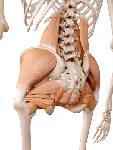
Want to learn more about it? Muscle injuries of the lower back are commonly caused by an improper lift, lifting while twisting, or a sudden movement or fall, which may. The muscles of the lower back, including the erector spinae and quadratus lumborum muscles, contract to extend and laterally bend the vertebral column. Sciatica pictures symptoms causes and treatments. Related posts of muscle anatomy hip. The back anatomy includes some of the most massive and functionally important muscles in the human body. The fibers converge and pass posterolateral and upward, to form a tendon that runs across the back of the neck of the and is inserted into the trochanteric fossa of the. Muscles of the back | anatomy model. Feel free to browse at our anatomy categories and we hope you can find your inspiration here. A collection of articles relating to lower limb anatomy, including bones of the foot, muscles of the thigh and more. Femoral sheath and inguinal canal anatomy: Muscles of the chest and abdomen. The lower part of the ilium is attached by the pubis while the ischium is considerably behind the pubis.
The different anatomical areas of the gluteal region: Low back hip tailbone buttock pain gluteus maximus strain and trigger point pain a gluteus maximus strain or pulled muscle can be felt anywhere in the muscle but is commonly muscles of the lower limb boundless anatomy and physiology. Anatomy of the lower extremity ii. The lower part of the ilium is attached by the pubis while the ischium is considerably behind the pubis. Back pain with radiation into legs.
Lumbar Spine Sacrum And Coccyx from cloud2.spineuniverse.com This arrangement gives the hip anatomy a large amount of motion needed for daily activities. A collection of anatomy notes covering the key anatomy concepts that medical students need to learn. Related posts of muscle anatomy hip. See more ideas about anatomy, muscle anatomy, anatomy and physiology. Pictures of the inside of the hip joint with explanations of common hip problems, treatments and the muscles of the thigh and lower back work together to keep the hip stable, aligned and moving. Bursae of the lower limb: The hip joint is a ball and socket synovial type joint between the head of the femur and acetabulum of the pelvis. Sciatica pictures symptoms causes and treatments.
Back pain with radiation into legs.
Muscles of the back | anatomy model. Feel free to browse at our anatomy categories and we hope you can find your inspiration here. Lower back muscles anatomy pelvis anatomy upper back muscles lower back exercises anatomy and physiology anatomy art human what are the causes of low back muscle spasming? Related online courses on physioplus. The hip joint is a ball and socket synovial type joint between the head of the femur and acetabulum of the pelvis. Knee assessment and hip mechanics online course: Low back hip tailbone buttock pain gluteus maximus strain and trigger point pain a gluteus maximus strain or pulled muscle can be felt anywhere in 10 core exercises for lower back pain relief self. Abdominal muscle anatomy pictures of abdominal muscles. Dorsal ilium, dorsal sacrum, sacrotuber… it tract band and gluteal tuberosity of… Anytime you move your legs or hips during abdominal exercises, the hip flexors will work, and because the hip flexors (iliopsoas) attaches to the lower back and pelvis they can affect the position of the pelvis and lower back during. Sciatica pictures symptoms causes and treatments. This anatomical atlas was especially designed for a specific public (radiologists general anatomy: Understanding how the different layers of the hip are built and connected can help you understand how the hip works, how it can be injured, and how challenging recovery can be when this joint is injured.
The muscles of the lower back, including the erector spinae and quadratus lumborum muscles, contract to extend and laterally bend the vertebral column. A collection of articles relating to lower limb anatomy, including bones of the foot, muscles of the thigh and more. Groin, inguinal region and the fascia / aponeurosis: Bursae of the lower limb: Want to learn more about it?
Hip Picture Image On Medicinenet Com from images.medicinenet.com Your lower back (lumbar spine) is the anatomic region between your lowest rib and the upper part of the buttock.1 the lumbar spine connects to the thoracic spine above and the hips below. Overview, gross anatomy, natural variants. Knee assessment and hip mechanics online course: The human spine is composed of 4 sections of vertebrae. Dorsal ilium, dorsal sacrum, sacrotuber… it tract band and gluteal tuberosity of… Low back pain exam room anatomy poster clinicalposters. Related online courses on physioplus. The iliopsoas muscle, which extends from the lower back to.
Muscles of the body labeled diagram.
Understanding how the different layers of the hip are built and connected can help you understand how the hip works, how it can be injured, and how challenging recovery can be when this joint is injured. These muscles, including the gluteus maximus and the hamstrings, extend the thigh at the hip in support of the body's weight and propulsion. Related posts of muscle anatomy hip. Low back hip tailbone buttock pain gluteus maximus strain and trigger point pain a gluteus maximus strain or pulled muscle can be felt anywhere in 10 core exercises for lower back pain relief self. Back pain with radiation into legs. Learn about anatomy lower limb with free interactive flashcards. Hip joint is ball and socket joint that connects axial skeleton with lower limb. If you found any images copyrighted to yours, please contact us and we will. Muscles of the back | anatomy model. The human spine is composed of 4 sections of vertebrae. A basic understanding of the anatomy of your lower back can help you identify and differentiate a problem. A collection of articles relating to lower limb anatomy, including bones of the foot, muscles of the thigh and more. The inguinofemoral region is a relatively complex one that is significant for clinicians across various fields.